Interdisciplinary projects break down the barriers between different fields enabling innovative initiatives to see the light of day. This type of microscope uses visible light view thicker larger specimens such as an insect in 3D.

Microscopy Types Applications Video Lesson Transcript Study Com
Funjournal Org
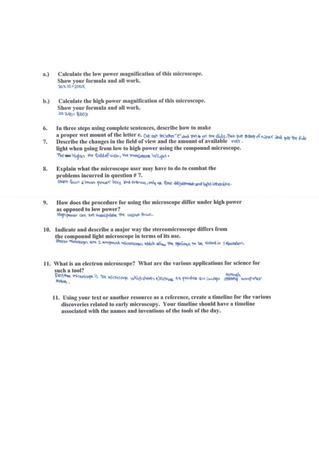
Microscope Lab
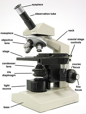
Use material from Section 181 of your text to label the condenser objective and ocular lenses in the.

Basic techniques in microscopy lab report. Be able to name the parts of the microscope and give the function of each part. This article gives an overview of metallography and metallic alloy characterization. The lab seeks to gain basic insights into problems that have wide socioeconomic impact from spine deformities in the young to falls in the elderly.
General Aseptic Techniques in Microbiology Laboratory. A response is required for each item marked. Scalable image processing techniques for quantitative analysis of volumetric biological images from light-sheet microscopy bioRxiv 2019 DOI.
When we talk about light in microscopy it is usually noted as wavelength even though photons the packets of energy that make light can act as both particles and waves. With the advent of electron microscopy its detailed cytology and molecular organization has been clearly established. These range from a form to fill in and submit before leaving the lab to a formal written report.
Biology is the science of life and life processes. A BIO 102 General Biological Sciences 3 Introduction to the major concepts in biology and a survey of the common structures of organisms including humans and their functions at the molecular cellular organismal and population levels. Fundamental research is carried out that seeks to understand how the brain coordinates and controls a myriad of muscles in human locomotion and how aging affects that control.
Park Systems a world leading manufacturer of Atomic Force Microscopes presents Park NX-Hybrid WLI the first fully integrated system that combines Atomic. What you need to be able to do on the exam after completing this lab exercise. The specimen is most often an ultrathin section less than 100 nm thick or a suspension on a grid.
Since you are viewing larger samples the magnification range of the dissecting microscope is lower than the compound light microscope. Basic microscopy deals with a diverse group of studies that help in research related to the biochemistry physiology cell biology ecology evolution and clinical aspects of microorganisms including the host response to these agents. However it is not without disadvantages and requires significant.
To observe prokaryotic cells and practice oil immersion techniques obtain a sample from the source plates or tubes on your lab bench Staphylococcus epidermidis Escherichia coli and Bacillus subtilis according to instructions below. Electron microscopy is a useful technique that allows us to view the microscopic structure of specimens at a high resolution. Through its five schools and three colleges EPFL explores many research fields and offers a wide range of training programmes.
We study both synthetic and biological materials. Fluorescence is the result of a three-stage process that occurs in certain molecules generally polyaromatic hydrocarbons or heterocycles called fluorophores or fluorescent dyes Figure 1A fluorescent probe is a fluorophore designed to respond to a specific stimulus or to localize within a specific region of a biological specimen. The techniques we use include light scattering optical microscopy rheology and microfluidics.
Another microscope that you will use in lab is a stereoscopic or a dissecting microscope. Lab reports can vary in length and format. From the lower leg of the grass frog Rana pipiens.
And Hang Lu Microfluidic chamber arrays for whole-organism behavior-based chemical screening Lab on a. Visible light or light that we can see with our eyes is usually in the range of 400700 nm and encompasses all colors in the rainbow with blues starting at around 400 nm and reds finishing. We are proud to announce that the Douglas Research Centre in partnership with three other McGill-affiliated research centres the RI-MUHC the Lady Davis Institute and the Centre de recherche en biologie structurale has been awarded new funding from the FRQS to improve career and professional development for research trainees.
Simple Staining of Bacteria Observing Under Oil Immersion. Metallography developed from the need to understand the influence of alloy microstructure on macroscopic properties. Scientific Report 2017 7 DOI101038srep46306.
The visible spectrum of light. Lab Exercise 2 Microscopy Cell Structure Textbook Reference. Gram Staining ProcedureProtocol.
In this laboratory we will look at the contractile properties of the. In addition it is an excellent experimental subject. Courses in Biological Sciences.
See Chapter 3 for Cell Structure. Different microscopy techniques are used to study the alloy microstructure ie microscale structure of grains phases inclusions etc. Be able to explain the proper usage and care of.
Flood air-dried heat-fixed smear of cells for 1 minute with crystal violet staining reagent. Our interests extend from fundamental physics to technological applications from basic materials questions to specific biological problems. The knowledge obtained is exploited for the design.
The Jackson Laboratory models and interprets genomic complexity integrates basic research with clinical application educates current and future scientists and empowers the global biomedical community by providing critical data tools and services. Your grade for the lab 1 report 1A and 1B combined will be the fraction of correct responses on a 50 point scale correct total x 50. ˌ b aɪ j ə ˈ r ɛ t ˈ b aɪ j ə ˌ r ɛ t test also known as Piotrowskis test is a chemical test used for detecting the presence of peptide bondsIn the presence of peptides a copperII ion forms mauve-colored coordination complexes in an alkaline solution.
Several variants on the test have been developed such as the BCA test and the Modified Lowry test. Please note that the quality of the smear too heavy or too light cell concentration will affect the Gram Stain results. It may include a variety of diverse activities such as the sequencing of DNA determining the rate of neuron activity or measuring plant diversity in a prairie.
Emphasis placed on principles of ecology inheritance evolution and physiology relevant to human society. An image is formed from the interaction of the electrons with the sample as the beam is transmitted through the specimen. The aseptic techniques control the opportunities for contamination of cultures by microorganisms from the environment or contamination of the environment by the microorganisms being handled.
Transmission electron microscopy TEM is a microscopy technique in which a beam of electrons is transmitted through a specimen to form an image.
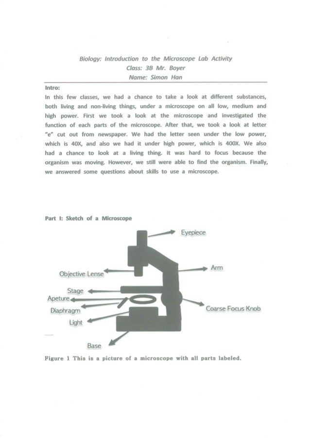
Microscope Lab
Lab 1 Principles And Use Of Microscope Ibg 102 Lab Report

Solved To Submit This Assignment Students Will Complete The Chegg Com
Funjournal Org
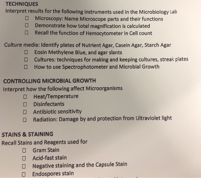
Solved Techniques Interpret Results For The Following Chegg Com
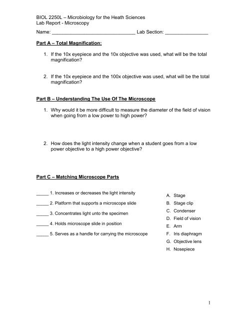
Microscopy Lab Report

Lab 1 Microscopy Lab Report 1 As 020 316 Cell Biology Lab Studocu
Microscope

